L'intelligenza artificiale nella diagnostica per immagini: applicazioni, vantaggi e implementazione

1 settembre 2026 10 minuti di lettura
Riassumere l'articolo con IA

Punti di forza

  • AI now handles detection, segmentation, triage, reconstruction, reporting, and prediction across radiology, cardiology, neurology, and MSK imaging.
  • Radiologists read less noise, patients get diagnosed earlier, providers move faster with the same headcount.
  • Without PACS, RIS, and EHR integration, even a great model is just an expensive demo.
  • Validation, human oversight, and ongoing monitoring are what keep AI from quietly drifting off course.

Lots of healthcare IT conference keynotes follow the same pattern by now. Someone opens with a slide of a friendly-looking robot in a white coat, then closes forty minutes later fielding a nervous question about whether the software is coming for the audience’s jobs. Somewhere between the robot slide and that question, the really useful part of the talk gets skipped over, and it’s rarely about robots.

AI for medical imaging, in the form that actually ships and gets used, is much less dramatic and much more interesting. It’s software that reads a chest CT before the radiologist opens the file and flags the three studies out of two hundred that need eyes on them first. It’s a model that measures a tumor the same way every single time, instead of the way a tired resident measures it at 6 p.m. on a Friday. None of it makes headlines. All of it changes a department’s workday.

This is a practical walkthrough of where AI in medical imaging is genuinely working, where it still trips over its own shoelaces, and what it takes to bring it into a hospital or imaging center without breaking anything important along the way.

What is artificial intelligence in medical imaging?

At its simplest, AI-based medical imaging means training software, mostly deep learning models built on convolutional neural networks or newer transformer architectures, to recognize patterns in scans the way a trained radiologist does. Feed a model enough labeled chest X-rays showing pneumonia, and it starts noticing the same visual cues a radiologist notices, just faster and without a coffee break.

The scope has grown well past finding a single tumor. Today’s tools touch nearly every stage of the imaging pipeline: acquisition, reconstruction, interpretation, reporting, and increasingly, predicting disease progression and patient outcomes. Some of this runs as machine learning baked directly into the scanner. Some of it runs as a separate layer that reads the DICOM file once the scan is done.

Regulators noticed the shift years before most healthcare organizations did. As of late 2025, radiology accounted for roughly 76% of every AI-enabled medical device cleared by the FDA in the last 30 years, more than every other clinical specialty combined. Imaging is where the data is richest, the patterns are visual enough for a neural network to learn well, and the clinical payoff is clearest, so it’s also where AI got its head start.

AI applications in medical imaging

AI does several different jobs depending on where it sits in the workflow. Here’s how AI is used in medical imaging once you get past the marketing slide.

Early abnormality detection

This is the application everyone pictures first. A model scans an image and flags anything that looks like it shouldn’t be there: a nodule, a fracture, a hemorrhage, a suspicious mass, etc. It stands to reason that the output isn’t a diagnosis. It’s just a second set of eyes that never gets tired or distracted, which is exactly what a visione artificiale model is good at, and exactly what a human reading their two hundredth chest X-ray of the shift is not.

Image segmentation and measurement

Segmentation draws the outline, marking exactly where a tumor, organ, or vessel is located on the image. It sounds unglamorous until you realize how much radiology depends on consistent measurement over time. A model that segments the same lesion consistently across follow-up scans removes a surprising amount of guesswork from treatment monitoring.

Worklist prioritization and clinical triage

Not every scan is equally urgent, but PACS (picture archiving and communication system) worklists have traditionally treated them that way: first in, first read. Triage models scan incoming studies and bump anything showing signs of stroke, pulmonary embolism, or intracranial bleeding to the top of the queue, sometimes within minutes of acquisition. The radiologist still makes the call. They just make it sooner.

Image reconstruction and enhancement

AI also works on the image itself before a human ever looks at it, cleaning up noise from low-dose CT scans, sharpening MRI images captured in less time than a standard protocol requires, or filling in gaps from motion artifacts. The upside is real: lower radiation doses, shorter scan times, and images that are easier to read.

Predictive and multimodal analytics

The newest wave combines imaging with other data types, lab results, genomics, and clinical notes to predict how a disease is likely to progress instead of just describing what’s visible today. It’s early, it’s promising, and it’s the direction a lot of the serious research money is currently pointed.

Want an imaging AI model built around your clinical workflow?

AI medical imaging use cases by clinical area

Applications are one way to slice this, but the clinical area is another. And it’s probably the one that matters more to whoever is signing off on the budget. Here are some concrete AI in medical imaging examples by specialty.

Cancer detection and treatment monitoring

Oncology imaging has one of the deepest AI track records, mostly because breast, lung, and prostate cancer screening produce enormous, well-labeled datasets. In one of the largest real-world trials to date, radiologists using AI-supported double reading in mammography screening detected 17.6% more cancers than radiologists working without it, across more than 460,000 women screened at 12 sites in Germany.

Neurological imaging

Stroke care runs on minutes, and AI triage tools built for CT and MRI now flag suspected large vessel occlusions and intracranial hemorrhage fast enough to shorten the time between scan and treatment decision. The same underlying tech also tracks white matter changes over time for conditions like multiple sclerosis, which used to require painstaking manual comparison between scans.

Cardiovascular imaging

Cardiac MRI and CT produce dense and motion-heavy data that’s genuinely hard for a human to measure consistently. AI tools now handle chamber volume calculations, calcium scoring, and coronary artery analysis with a level of repeatability that manual measurements rarely achieve, since two cardiologists measuring the same heart by hand won’t always land on the exact same number.

Musculoskeletal imaging

Fracture detection on X-ray, especially subtle fractures that are easy to miss in a busy emergency department, is one of the quieter AI success stories. Bone age assessment and osteoarthritis grading have followed the same pattern: tasks that were always somewhat subjective now have a consistent second opinion attached.

Benefits of AI in medical imaging and diagnostics

The benefits of AI in medical imaging land differently depending on who’s asking, so it’s worth breaking them apart by audience instead of pretending everyone wants the same thing.

Radiologi

  • Fewer routine, low-value reads, and more time on complex cases that need a specialist’s judgment
  • A consistent second read that catches fatigue-driven misses, particularly late in a shift
  • Faster, partially pre-filled reports that cut down on repetitive dictation

Pazienti

  • Earlier detection for time-sensitive conditions like stroke and certain cancers
  • Lower radiation exposure through AI-enhanced low-dose CT imaging 
  • Shorter waits for results, since triaged, urgent studies get read first

Fornitori di assistenza sanitaria

  • Better throughput from the same radiologist headcount, which matters given how hard imaging specialists are to hire
  • Lower long-term costs from fewer repeat scans and fewer missed findings that turn into bigger problems later
  • Data that supports quality reporting and accreditation, since most AI tools log exactly what they found and when

Want to see what AI-ready healthcare software looks like?

Challenges of AI-based medical imaging

None of the above happens automatically, and pretending otherwise is how AI pilots quietly die in a drawer somewhere. Here’s what tends to get in the way.

Data quality and annotation

A model is only as good as the labels it learned from, and medical image annotation is slow, expensive, and requires actual clinical expertise. Inconsistent labeling between institutions is one of the most common reasons a model performs beautifully in testing and disappointingly in production.

Bias and limited generalization

Models trained mostly on one population, one scanner brand, or one hospital’s imaging protocols tend to underperform everywhere else. A model that learned breast density patterns from one demographic doesn’t automatically transfer to a different one.

False positives and false negatives

Every threshold is a tradeoff. Set it too sensitive and radiologists drown in false alarms, which breeds the exact alert fatigue the tool was supposed to prevent. Push too far toward specificity and it starts missing real findings. There’s no setting that makes this tradeoff disappear, only settings that suit a particular clinical context better or worse.

Explainability and clinical trust

A model that says “cancer, 87% confidence” without showing why is a hard sell to a radiologist who’s legally and professionally accountable for the final call. Tools that highlight the actual pixels driving a prediction earn trust faster than ones that don’t, mostly because they let a clinician sanity check an AI medical imaging diagnosis.

Privacy and cybersecurity

Medical images carry protected health information, and AI pipelines that move that data between scanners, storage, cloud inference, and reporting systems multiply the places where something can go wrong. Encryption in transit and at rest, strict access controls, and audit logging are what keep a breach from becoming a regulatory disaster.

Regulation and accountability

Who’s responsible when an AI-assisted read misses something? Regulatory frameworks are still catching up to that question, and clearance from a body like the FDA covers the software’s performance, not the legal and clinical accountability structure around how it’s used.

Model drift and monitoring

A model validated on last year’s scanner models and patient mix can quietly degrade as equipment, protocols, and populations shift, a problem known as drift. Without ongoing performance monitoring, that degradation is invisible until someone notices the model has been wrong for months. This is exactly the kind of thing solid MLOps practices are built to catch before it becomes a clinical problem.

How AI fits into medical imaging workflows

Getting a model to work in a research paper and getting it to work inside a real radiology department are two very different projects. Here’s the shape of the second one.

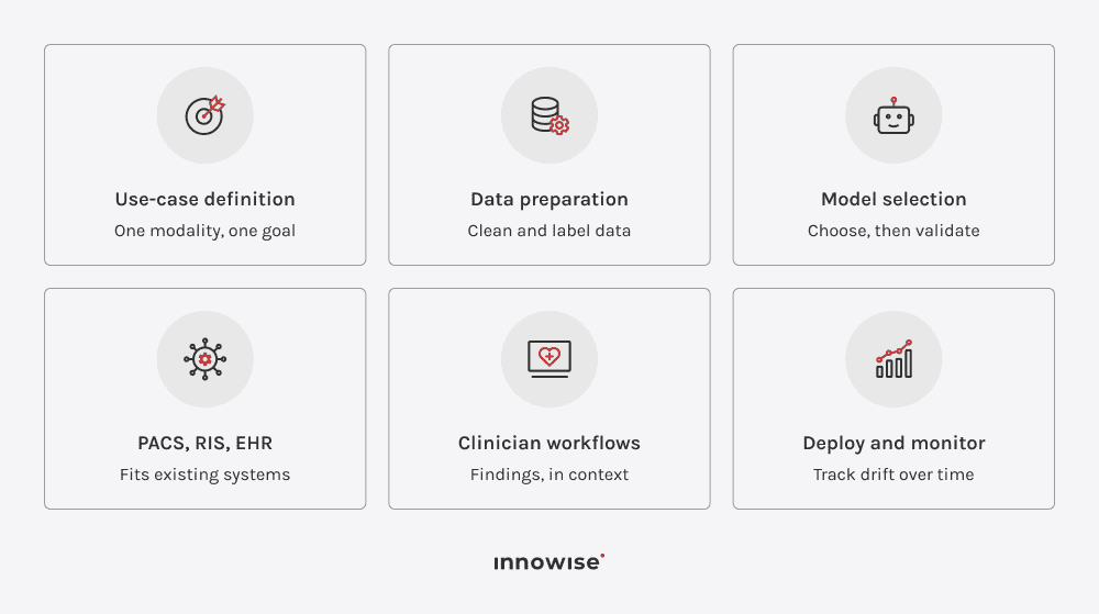
Clinical use-case definition

Every successful deployment starts narrow: one modality, one condition, one clearly defined clinical question. “AI for radiology” is not a use case. “Flag suspected pulmonary embolism on chest CT within five minutes of acquisition” is.

Preparazione dei dati

This is where most of the real work hides: cleaning, de-identifying, and annotating imaging data to a standard the model can learn from. The next challenge is making sure the data reflects the patient population and equipment the model will eventually run on.

Model selection and validation

Off-the-shelf model, fine-tuned foundation model, or fully custom build? The right answer depends on how common the clinical task is and how much your patient population differs from what existing models were trained on. We’ll come back to that in the next section.

PACS, RIS, and EHR integration

A model that can’t receive studies from PACS, write findings back to RIS, and surface results inside the EHR a clinician is already using isn’t a clinical tool but a research project with good intentions. Integrazione tra EHR ed EMR work is unglamorous and absolutely essential, because nobody wants to open a third application just to see what the AI found.

Clinician-facing workflows

The interface matters almost as much as the model underneath it. AI findings need to appear within the tools radiologists already use and be presented in a format that fits their clinical workflow, with simple options to accept, reject, or override the AI’s suggestions.

Implementazione e monitoraggio

Surely, go-live isn’t the finish line. Ongoing monitoring tracks accuracy against real outcomes, watches for drift, and catches the moment a firmware update on the scanner quietly changes the images the model is seeing.

In-house build, off-the-shelf buy, or a delivery partner?

Sooner or later, every AI medical imaging technology plan hits the same crossroads, and somebody in the room says the word “roadmap” with more confidence than the situation deserves. You can build the thing yourselves, buy something that already exists, or hire people who have already made the expensive mistakes on someone else’s budget. None of these three is correct in the abstract. One of them is correct for your team, your deadline, and how much risk your legal department can stomach before lunch.

ApproccioSpeed to deployCosto inizialeClinical customizationManutenzione a lungo termine
Build in-houseSlow: months to yearsHigh, requires a dedicated ML teamMolto altoFalls entirely on your team
Buy off-the-shelfFast: weeksLower upfront, ongoing license feesLimited to what the vendor supportsHandled by the vendor
Partner with a dev teamModerate: weeks to a few months, a yearModerate, scoped to the projectAltoShared or handed off after stabilization

Technology requirements

Good intentions don’t pass a security audit, unfortunately. Before an AI/ML medical imaging pipeline gets anywhere near an actual patient, a fairly unglamorous stack has to already be sitting underneath it, quietly doing its job so nobody has to think about it.

  • DICOM and DICOMweb, for moving and displaying imaging data in the format radiology equipment already speaks
  • PACS and RIS, the systems that store, route, and track imaging studies
  • HL7 and FHIR, for exchanging structured clinical data with the EHR
  • Cloud, edge, and GPU infrastructure, sized for inference speed, not just training
  • MLOps and model registries, to version, monitor, and roll back models safely
  • Identity and access management, controlling exactly who can see what
  • Encryption, audit logs, and monitoring, covering data in transit, at rest, and in use

How to implement AI in medical imaging

Here’s roughly the order we walk clients through, assembled from projects where the AI imaging solutions made it past the pilot and into an ordinary Tuesday.

  1. Pick the clinical problem and write down measurable KPIs before anyone touches a line of code. A vague goal is how you end up with a model nobody can actually use.
  2. Take an honest look at your existing imaging data and infrastructure, gaps included. Most delays trace back to a data quality issue nobody wanted to mention in the kickoff meeting.
  3. Decide whether to build, buy, or partner, using the comparison above and, more importantly, your team’s actual bandwidth, not its optimistic bandwidth.
  4. Validate the model against data that looks like your real patient population. The vendor’s demo dataset is not that.
  5. Integrate the tool into PACS, RIS, and EHR workflows so the findings show up where clinicians already are, not in a separate tab they’ll forget exists.
  6. Run a controlled pilot with a defined group of radiologists and a way to measure results against a real baseline, not a feeling.
  7. Train radiologists and operational staff properly. A tool nobody trusts gets quietly ignored, no matter how impressive its accuracy numbers looked on the pitch.
  8. Keep monitoring clinical, technical, and business results well past the pilot window, because that’s usually where the real story shows up.
  9. Only expand to new sites, modalities, or use cases once the first deployment has proven it can behave itself.

Not sure whether to build, buy, or partner for your imaging AI project?

The future of AI in medical imaging

A handful of directions keep turning up in serious research and product roadmaps often enough that they’re worth watching, not filed away as vendor slideware. This is roughly where AI is transforming medical imaging once teams outgrow the current toolkit.

  • Multimodal and foundation models, trained across many imaging types and text data at once instead of one narrow task
  • Generative reporting support, drafting structured findings a radiologist reviews and edits instead of writing from scratch
  • Longitudinal patient analysis, tracking how a patient’s imaging changes across years, not just comparing two scans side by side
  • Personalized imaging protocols, adjusting scan parameters to an individual patient instead of one fixed setting for everyone
  • Federated learning, training models across multiple hospitals’ data without the raw patient data ever leaving the building it started in
  • AI agents for radiology workflow coordination, handling scheduling, triage, and follow-up tracking around the actual image interpretation

How Innowise supports AI medical imaging projects

Our work here spans the AI healthcare medical imaging stack end to end. On one side, visione artificiale models trained on real clinical imaging data. On the less glamorous side, the PACS, RIS, and EHR integration work that decides whether any of it gets used. HIPAA, GDPR, and MDR compliance aren’t bolted on at the end. They’re part of the build from the first sprint, and our ISO 13485 certification covers exactly the kind of medical device software this touches. Our healthcare case studies give a fairer picture of what that’s looked like in practice than anything I could sum up here.

Conclusione

AI won’t win against a broken PACS integration, and it won’t wait around for a perfect one either. Somewhere between the pilot program and the messy reality of hospital IT, radiology departments are already reading images differently than they did five years ago. Whether that shift feels revolutionary or just quietly useful has less to do with the algorithm than with who was willing to do the often-overlooked integration work first.

FAQ

Un po’ di tutto, a dire il vero: rilevamento delle anomalie, segmentazione e misurazione delle immagini, triage delle liste di lavoro, ricostruzione delle immagini, refertazione strutturata e analisi predittiva, in una vasta gamma di modalità quali radiografia, TAC, risonanza magnetica ed ecografia.

Raggi X, TAC, risonanza magnetica, ecografia, mammografia, PET e, sempre più spesso, immagini patologiche di vetrini interi. I raggi X e la TAC vantano ancora la tradizione più lunga e consolidata.

Nella maggior parte dei casi, sì. Gli strumenti clinici basati sull’intelligenza artificiale sono generalmente progettati per integrarsi con i sistemi PACS e RIS esistenti tramite standard quali DICOM, DICOMweb, HL7 V2 e FHIR, anche se il grado effettivo di integrazione dipende ancora in larga misura dal fornitore.

Ciò dipende in larga misura dal tipo di compito e dal grado di rigore con cui il modello è stato convalidato, quindi è bene considerare con una certa cautela qualsiasi dato isolato relativo all’accuratezza. Strumenti ben convalidati in ambiti come lo screening mammografico hanno dimostrato miglioramenti concreti nei tassi di rilevamento rispetto alla lettura non assistita, insieme a compromessi reali nei tassi di falsi positivi che vale la pena valutare.

Non esiste una cifra magica. Dipende dalla complessità del compito clinico, ma i modelli di imaging convalidati e approvati dalla FDA hanno storicamente utilizzato un numero di casi clinici che va da poche centinaia a diverse migliaia per la validazione clinica.

Negli Stati Uniti, principalmente l'autorizzazione della FDA attraverso procedure quali la 510(k), la De Novo o la PMA, oltre alla legge HIPAA in materia di protezione dei dati. Nell'Unione Europea, si tratta del Regolamento sui dispositivi medici (MDR), del GDPR e della legge UE sull'intelligenza artificiale.

La risposta breve è no. Gli strumenti attualmente disponibili sono approvati come ausili di lettura di supporto o complementari, non come lettori autonomi, quindi la decisione clinica finale spetta comunque al radiologo, non a un modello.

Onestamente, le variabili sono troppe per poter indicare una cifra precisa. Acquistare una soluzione già pronta, svilupparne una su misura o collaborare con un team di sviluppo comportano costi molto diversi tra loro, così come la complessità dell’integrazione e il numero di siti coinvolti. Una consulenza mirata ti fornirà una cifra concreta molto più rapidamente di quanto non possa fare una semplice stima.

Mostra tutto

Healthcare IT Consultant

Aleh colma il divario tra le esigenze cliniche e l'esecuzione tecnica. Applica una profonda conoscenza del settore per garantire che i sistemi MedTech non siano solo conformi, ma anche sufficientemente affidabili da avere un impatto misurabile nell'assistenza sanitaria reale.

Indice dei contenuti

    Contattaci

    Prenota una chiamata oppure compila il modulo qui sotto e ti ricontatteremo non appena avremo elaborato la richiesta.

    Inviaci un messaggio vocale
    Allega i documenti
    Carica il file

    È possibile allegare 1 file di dimensioni massime di 2 MB. Formati di file validi: pdf, jpg, jpeg, png.

    Facendo clic su «Invia», acconsenti al trattamento dei tuoi dati personali da parte di Innowise in conformità con la nostra Informativa sulla privacy per fornirti informazioni pertinenti. Inviando il tuo numero di telefono, accetti che possiamo contattarti tramite chiamate vocali, SMS e app di messaggistica. Potrebbero essere applicati costi per chiamate, messaggi e traffico dati.

    Puoi anche inviarci la tua richiesta
    a contact@innowise.com
    Cosa succede dopo?
    1

    Una volta ricevuta e presa in carico la richiesta, ti ricontatteremo per approfondire le esigenze del progetto e firmare un NDA per tutelare la riservatezza.

    2

    Dopo aver chiarito esigenze, obiettivi e aspettative, il nostro team elabora una proposta che definisce le attività previste, la composizione del team, le tempistiche e i costi stimati.

    3

    Fisseremo un incontro per discutere la proposta e definire tutti i dettagli.

    4

    Infine, firmeremo il contratto e inizieremo subito a lavorare sul tuo progetto.

    Altri servizi che copriamo

    arrow